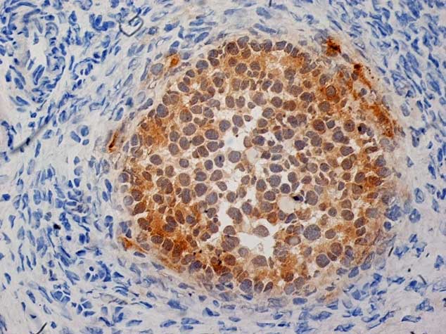
Alpha inhibin
It appears that your browser has cookies disabled.
Inhibin is a peptide hormone that is produced by ovarian granulosa cells which inhibits the release of Follicle-Stimulating Hormone FSH. The Inhibin alpha antibody subunit is expressed in a wide range of human tissues outside the reproductive axis such as prostate, brain, adrenal, as well as in the granulosa cells of the ovary, Sertoli cells of the testis and various cells of the fetoplacental unit. Inhibin may be used as a differential marker for adrenocortical tumors, placenta and gestational trophoblastic lesions and sex cord stromal tumors. Normal testis or normal ovary, adrenal gland. Groome, N G et al Monoclonal and polyclonal antibodies reactive with the amino terminal sequence of the alpha subunit of human 32K inhibin. Hybridoma 9 1 : 2.
Alpha inhibin
Federal government websites often end in. The site is secure. As a result of its expression in corresponding normal cell types, inhibin alpha INHA is used as an immunohistochemical marker for adrenocortical neoplasms and testicular or ovarian sex cord stromal tumors. However, other tumors can also express INHA. To comprehensively determine INHA expression in cancer, a tissue microarray containing 15, samples from different tumor types and subtypes was analyzed by immunohistochemistry. In summary, these data support the use of INHA antibodies for detecting sex cord stromal tumors, granular cell tumors, and adrenocortical neoplasms. Since INHA can also be found in other tumor entities, INHA immunohistochemistry should only be considered as a part of any panel for the distinction of tumor entities. The inhibin alpha subunit protein INHA is a member of the TGF-beta transforming growth factor-beta superfamily encoded by a gene located at 2q35 [ 1 , 2 , 3 ]. It combines with the A and B type proteins of the inhibin beta subunits to form inhibin protein complexes that negatively regulate the secretion of follicle-stimulating hormone FSH from the pituitary gland [ 4 , 5 , 6 ]. Inhibin has also been suggested to inhibit gonadal stromal cell proliferation and to possess a tumor suppressive activity [ 4 ]. Among normal tissues, INHA staining is found in adreno-cortical cells, Sertoli and Leydig cells of the testis, and the placenta [ 7 ]. Accordingly, inhibin alpha is currently used as an immunohistochemical marker for adrenocortical tumors and sex cord stromal tumors of the testis and the ovary [ 7 , 8 , 9 ].
Inhibin alpha is a mouse monoclonal antibody derived from cell culture supernatant that is concentrated, alpha inhibin, dialyzed, filter sterilized and diluted in buffer pH 7. Board review style answer 2.
Check out our latest pathology themed Wordle here! Updated every Monday. Editorial Board Member: Christian M. Page views in 13, Cite this page: Wirth P.
It appears that your browser has cookies disabled. The website requires your browser to enable cookies in order to login. Don't have a login? Register today to view list prices and apply for an account to purchase online! Inhibins and activins play a role in the regulation of pituitary follicle stimulating hormone FSH secretion. The actions of inhibins and activins are thought to oppose one another, with inhibins suppressing FSH secretion and activins stimulating FSH secretion. Inhibins are secreted by granulosa cells in female follicles and Sertoli cells of the testis in the male.
Alpha inhibin
Check out our latest pathology themed Wordle here! Updated every Monday. Editorial Board Member: Christian M.
Alebrijes dibujos
Lim D. A comparison of A and inhibin reactivity in adrenal cortical tumors: Distinction from hepatocellular carcinoma and renal tumors. Availability Catalog No. Successfully added to cart added to cart. Download SDS Sheet. Board review style question 1. All work was carried out in compliance with the Helsinki Declaration. Non-invasive papillary urothelial carcinoma, pTa G2 low grade. Download Data Sheet. Isolation and partial characterization of a Mr 32, protein with inhibin activity from porcine follicular fluid. More than 12, tumors were successfully analyzed in this study. IVD - CE. Microscopic histologic description. Sakr S.
Request Quote Add To Cart.
Hybridoma 9 1 : 2. Table S1: List of the references and raw data used to create Figure 3. Facebook page opens in new window Twitter page opens in new window Instagram page opens in new window. Due to the rareness of immunohistochemically detectable INHA expression in most cancer types, we were only able to compare INHA immunostaining data with available clinical data in neuroendocrine tumors, clear cell renal cell carcinoma, and colorectal adenocarcinoma. Clone: R1. Detailed histopathological data were available for 2, colorectal adenocarcinomas, neuroendocrine tumors, and clear cell renal cell carcinomas. The images A—F and G—M are from consecutive tissue sections. Related products. Clinical features. E Mayo K. This website is intended for pathologists and laboratory personnel but not for patients.


Excuse for that I interfere � But this theme is very close to me. I can help with the answer.