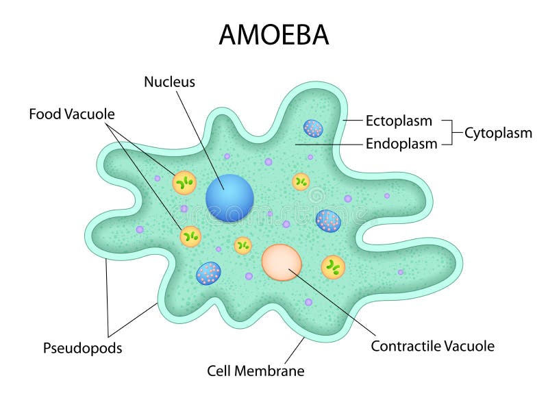
Amoeba ka diagram
Amoeba is a unicellular organism that has the ability to change its shape.
Amoeba unicellular animal with pseudopods that lives in fresh or saltwater. Anatomy of an amoeba. Vector illustration for medical, educational and science use. Amoeba binary fission infographic. Vector illustration of reproduction of simplest bacteria.
Amoeba ka diagram
.
Infusoria isolated on white. One of the first reports referencing amoebas dates can be traced back to 18th century.
.
Amoebas are unicellular eukaryotic organisms classified in the Kingdom Protista. Amoebas are amorphous and appear as jelly-like blobs as they move about. These microscopic protozoa move by changing their shape, exhibiting a unique type of crawling motion that has come to be known as amoeboid movement. Amoebas make their homes in salt water and freshwater aquatic environments , wet soils, and some parasitic amoebas inhabit animals and humans. Amoebas are simple in form consisting of cytoplasm surrounded by a cell membrane. The outer portion of the cytoplasm ectoplasm is clear and gel-like, while the inner portion of the cytoplasm endoplasm is granular and contains organelles , such as a nuclei , mitochondria , and vacuoles. Some vacuoles digest food, while others expel excess water and waste from the cell through the plasma membrane.
Amoeba ka diagram
Amoeba proteus is a unicellular organism widely distributed in ponds, lakes, freshwater pools and slow streams. Normally it is found creeping, feeding upon algae, bacteria etc. Under the microscope, it appears as irregular, jelly-like tiny mass of hyaline protoplasm. Amoeba has no fixed shape and the outline of body continues changing due to formation of small finger like outgrowths called pseudopodia fig. Pseudopodia are temporary finger like projections with blunt rounded tips which are constantly being given out or withdrawn by the body. Many pseudopodia are formed simultaneously. Amoeba exhibits movement by the pseudopodia. It also helps in food capture.
Gtowizard
Types of infectious agents. Vector diagram for educational, science, and biological use. Infusoria isolated on white. Search by image or video. Educational labeled scheme with bacteria inner parts as zoology study vector illustration. Amoeba labeled vector illustration. Typically, most amoebas are characterized by the following features:. Microbe is an organism of microscopic size, single-celled form or as a colony of cells. Tongue Function. Paramecium microscopic closeup structure with anatomical outline
FAQ Contact. Reimagine New Create image variations with AI. Pikaso Sketch to image with real-time AI drawing.
Educational single cell animal structure scheme with ectoplasm, nucleus, mitochondrion, pseudopod, lysosome and contractile vacuole. Labeled cell reproduction division stages scheme. What Is Endangered Species. Gut brain connection, dysbiosis and microbiome. Ribosome,cell wall,plasmid and chromosome copying steps. Vector illustration for educational and science use. Biology science educational information. Vector diagram for educational, science, and biological use. Vector illustration. Browse millions of high-quality stock photos, illustrations, and videos. Structure Infusorian of the shoeshoe type or Paramecium caudatum. Euglena outline vector illustration, science educated art. Colon and bowel vector illustration. Black silhouette of Chlamydomonas cell.


Rather good idea
The matchless answer ;)
Where I can read about it?