Emt epithelial mesenchymal transition
Federal government websites often end in.
Federal government websites often end in. The site is secure. The origins of the mesenchymal cells participating in tissue repair and pathological processes, notably tissue fibrosis, tumor invasiveness, and metastasis, are poorly understood. However, emerging evidence suggests that epithelial-mesenchymal transitions EMTs represent one important source of these cells. As we discuss here, processes similar to the EMTs associated with embryo implantation, embryogenesis, and organ development are appropriated and subverted by chronically inflamed tissues and neoplasias. The identification of the signaling pathways that lead to activation of EMT programs during these disease processes is providing new insights into the plasticity of cellular phenotypes and possible therapeutic interventions.
Emt epithelial mesenchymal transition
Thank you for visiting nature. You are using a browser version with limited support for CSS. To obtain the best experience, we recommend you use a more up to date browser or turn off compatibility mode in Internet Explorer. In the meantime, to ensure continued support, we are displaying the site without styles and JavaScript. An Author Correction to this article was published on 15 October Epithelial—mesenchymal transition EMT encompasses dynamic changes in cellular organization from epithelial to mesenchymal phenotypes, which leads to functional changes in cell migration and invasion. EMT occurs in a diverse range of physiological and pathological conditions and is driven by a conserved set of inducing signals, transcriptional regulators and downstream effectors. With over 5, publications indexed by Web of Science in alone, research on EMT is expanding rapidly. This growing interest warrants the need for a consensus among researchers when referring to and undertaking research on EMT. We trust that these guidelines will help to reduce misunderstanding and misinterpretation of research data generated in various experimental models and to promote cross-disciplinary collaboration to identify and address key open questions in this research field. While recognizing the importance of maintaining diversity in experimental approaches and conceptual frameworks, we emphasize that lasting contributions of EMT research to increasing our understanding of developmental processes and combatting cancer and other diseases depend on the adoption of a unified terminology to describe EMT.
Thus, elucidating EMT in 3D matrix requires careful consideration of tissue architecture and microenvironmental cues. Fibroblast growth factor signalling controls successive cell behaviours during mesoderm layer formation in Drosophila.
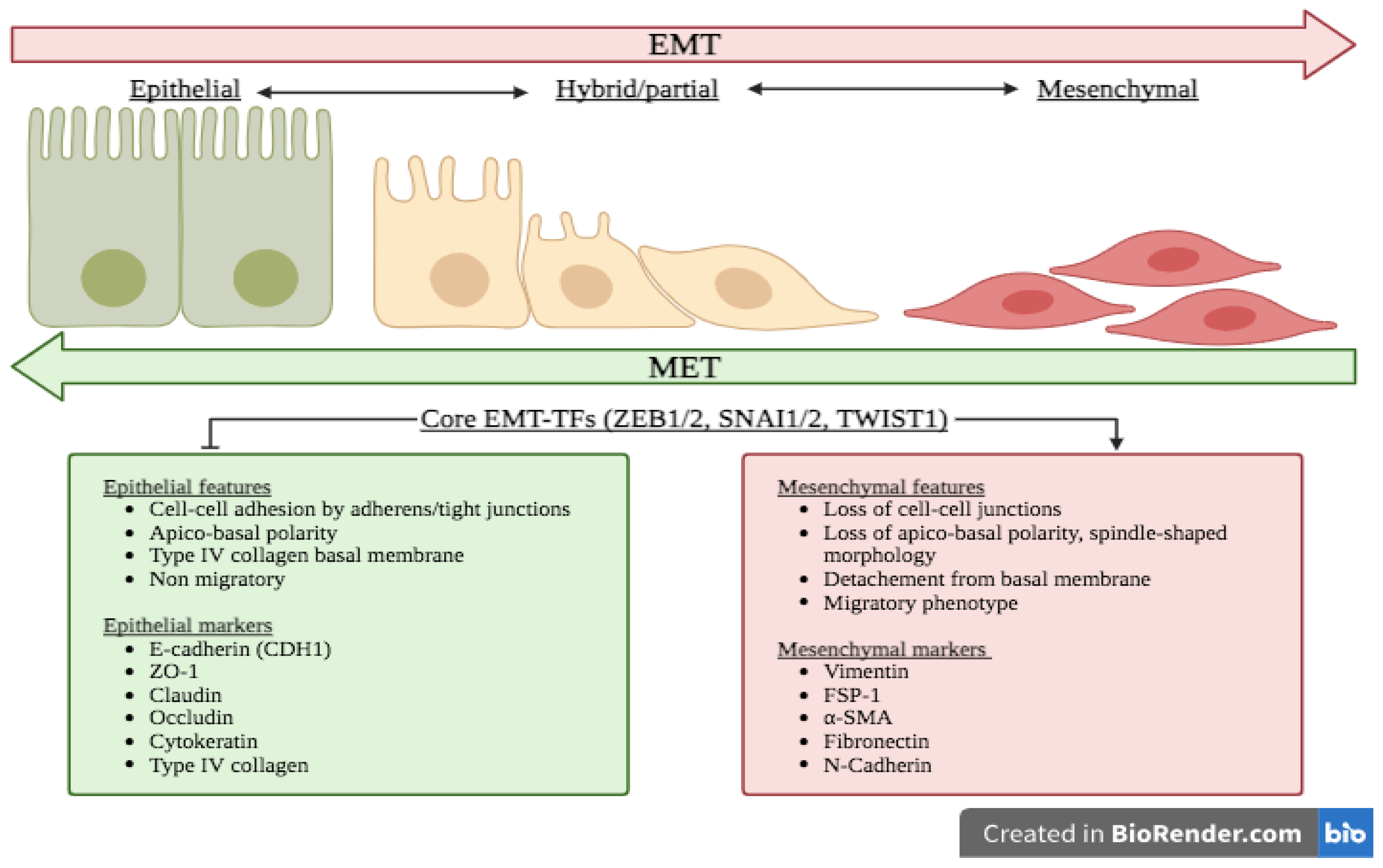
Abstract Epithelial-mesenchymal transition EMT and its reversal, mesenchymal-epithelial transition MET , are essential morphological processes during development and in the regulation of stem cell pluripotency, yet these processes are also activated in pathological contexts, such as in fibrosis and cancer progression. Multi-component signaling pathways cooperate in initiation of EMT and MET programs, via transcriptional, post-transcriptional, translational, and post-translational regulation. EMT is required for tissue regeneration and normal embryonic development as it enables epithelial cells to acquire the mesenchymal phenotype, conferring them migratory and dynamic properties towards forming threedimensional structures during gastrulation and organ formation. Uncontrolled activation of such phenomenon and the pathways signaling EMT events in adult life, leads to cancer growth and orchestrated by signaling interactions from the microenvironment, epithelial tumor cells with enhanced polarity, become invasive and rapidly metastasize to distant sites. Loss of epithelial markers E-cadherin and gain of mesenchymal markers N-cadherin , at the leading edge of solid tumors is associated with progression to metastasis.
Thank you for visiting nature. You are using a browser version with limited support for CSS. To obtain the best experience, we recommend you use a more up to date browser or turn off compatibility mode in Internet Explorer. In the meantime, to ensure continued support, we are displaying the site without styles and JavaScript. This is a preview of subscription content, access via your institution. Acloque, H. Epithelial-mesenchymal transitions: the importance of changing cell state in development and disease.
Emt epithelial mesenchymal transition
Federal government websites often end in. The site is secure. The origins of the mesenchymal cells participating in tissue repair and pathological processes, notably tissue fibrosis, tumor invasiveness, and metastasis, are poorly understood. However, emerging evidence suggests that epithelial-mesenchymal transitions EMTs represent one important source of these cells. As we discuss here, processes similar to the EMTs associated with embryo implantation, embryogenesis, and organ development are appropriated and subverted by chronically inflamed tissues and neoplasias. The identification of the signaling pathways that lead to activation of EMT programs during these disease processes is providing new insights into the plasticity of cellular phenotypes and possible therapeutic interventions. An epithelial-mesenchymal transition EMT is a biologic process that allows a polarized epithelial cell, which normally interacts with basement membrane via its basal surface, to undergo multiple biochemical changes that enable it to assume a mesenchymal cell phenotype, which includes enhanced migratory capacity, invasiveness, elevated resistance to apoptosis, and greatly increased production of ECM components 1. The completion of an EMT is signaled by the degradation of underlying basement membrane and the formation of a mesenchymal cell that can migrate away from the epithelial layer in which it originated.
Loungefly sale uk
Challenges in long-term imaging and quantification of single-cell dynamics. F-actin polymerization can be spatially coordinated along the cellular periphery; F-actin polymerization in a bundled state drives localized cellular protrusions i. The relationship of EMT to growth factors was found in Aiello, N. Activation of developmental EMT has been found to follow a defined sequence of events Fig. In particular, mammary epithelial cells at the core of a multicellular cluster were smaller and stiffer, while cells at the periphery were larger and softer [ ]. The expression of Snail and E-cadherin correlates inversely with the prognosis of patients suffering from breast cancer or oral squamous cell carcinoma , Epithelial cells come under the influence of these signaling molecules and, acting together with the inflammatory cells, induce basement membrane damage and focal degradation of type IV collagen and laminin Oft M. This process can also include an intermediate step of matrix remodeling via localized proteolysis e. Motility-limited aggregation of mammary epithelial cells into fractal-like clusters. Kalluri R. Studies using cell lines, developmental systems and cancer models have revealed a diversity of EMT-induced phenotypes and have highlighted remarkable complexity in the execution and regulation of EMT. Epithelial—mesenchymal transition EMT encompasses dynamic changes in cellular organization from epithelial to mesenchymal phenotypes, which leads to functional changes in cell migration and invasion.
Federal government websites often end in. The site is secure.
This initial recruitment of epithelial cells into an EMT can be inhibited by blocking the expression of MMP-9 through the disruption of tissue plasminogen activator tPA Nakaya, Y. Bridging from single to collective cell migration: a review of models and links to experiments. Oncogene 19 , — Cancer Cell. EMT is associated with the dynamic acquisition of an elongated, migratory phenotype, which is mediated by a redistribution of cell—matrix adhesions [ 22 ]. While not yet documented, it is plausible that many of the molecular regulators of EMTs also play critical roles in orchestrating EndMTs. Quantifying forces in cell biology. Although EMT has largely been studied at the level of altered gene transcription or posttranslational modifications, it is becoming increasingly clear that alternative splicing AS processes provide an additional layer of gene regulation that is critical in shaping the EMT process, particularly in cancer progression Tavanez and Valcarcel Typically, single cells formed compact acini on soft substrates, but disseminated as elongated mesenchymal cells on stiffened substrates. Extracellular matrix stiffness and composition jointly regulate the induction of malignant phenotypes in mammary epithelium. Obtaining quantitative EMT marker measurements should always be coupled with cellular and functional analyses of EMT status, as described above. Subsequent investigations revealed that CD8 T cells were required for outgrowth of the neu-negative mesenchymal variants suggesting local induction of EMT rather than selection Santisteban et al. Loss of E-cadherin is considered to be a fundamental event in EMT.


0 thoughts on “Emt epithelial mesenchymal transition”