Fluorodeoxyglucose positron emission tomography
The use of fluorodeoxyglucose positron emission tomography FDG PET scan technology in the management of head and neck cancers continues to increase.
Positron emission tomography PET [1] is a functional imaging technique that uses radioactive substances known as radiotracers to visualize and measure changes in metabolic processes , and in other physiological activities including blood flow , regional chemical composition, and absorption. Different tracers are used for various imaging purposes, depending on the target process within the body. PET is a common imaging technique , a medical scintillography technique used in nuclear medicine. A radiopharmaceutical — a radioisotope attached to a drug — is injected into the body as a tracer. When the radiopharmaceutical undergoes beta plus decay , a positron is emitted, and when the positron interacts with an ordinary electron, the two particles annihilate and two gamma rays are emitted in opposite directions.
Fluorodeoxyglucose positron emission tomography
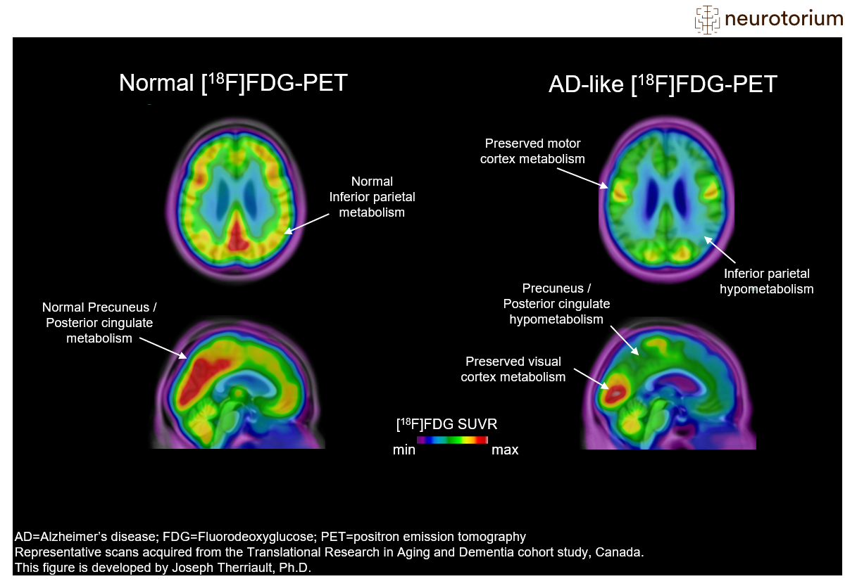
Federal government websites often end in. Before sharing sensitive information, make sure you're on a federal government site. The site is secure. NCBI Bookshelf. Muhammad A. Ashraf ; Amandeep Goyal. Authors Muhammad A. Ashraf 1 ; Amandeep Goyal 2. Fludeoxyglucose F18 is a radioactive tracer that acts as a glucose analog and is used for diagnostic purposes in conjunction with positron-emitting tomography PET to localize the tissues with altered glucose metabolism. It does not have therapeutic use. Altered glucose metabolism has implications for malignancies, epilepsy, myocardial ischemia, inflammatory conditions, and Alzheimer disease. PET scan uses radiotracers injected into the patient before the scan to visualize the blood flow and metabolic and biochemical activities in diseased and healthy tissues. FDG is a glucose analog that tends to accumulate in the tissue with high glucose demand, like tumors and inflammatory cells.
Dedicated PET scanners consist of multiple detectors that are arranged in a partial or full ring; the detection sensitivity of a partial ring scanner is less than that of a full ring scanner.
Yee C. Ung, Donna E. Maziak, Jessica A. Vanderveen, Christopher A. Lung cancer is the leading cause of cancer-related death in industrialized countries. The overall mortality rate for lung cancer is high, and early diagnosis provides the best chance for survival.
Federal government websites often end in. The site is secure. Preview improvements coming to the PMC website in October Learn More or Try it out now. Infections involving the heart are becoming increasingly common, and a timely diagnosis of utmost importance, despite its challenges. This review also functions to highlight the need for a standardized acquisition protocol. Infections of the heart primarily manifest as endocarditis, which affects 7. Prosthetic materials, including valve replacements, grafts, implantable devices, and related materials, such as leads, can lead to the development of endocarditis [ 2 ]. Moreover, cardiac implantable electronic device CIED infections occur in approximately 1. The diagnosis of such infections can be difficult, and the clinical presentation, as well as microbiological and imaging approaches have been used to reach an accurate diagnosis.
Fluorodeoxyglucose positron emission tomography
Background: Rhabdomyosarcoma RMS is the most common paediatric soft-tissue sarcoma and can emerge throughout the whole body. For patients with newly diagnosed RMS, prognosis for survival depends on multiple factors such as histology, tumour site, and extent of the disease. Patients with metastatic disease at diagnosis have impaired prognosis compared to those with localised disease. Appropriate staging at diagnosis therefore plays an important role in choosing the right treatment regimen for an individual patient. Fluorinefluorodeoxyglucose 18 F-FDG positron emission tomography PET is a functional molecular imaging technique that uses the increased glycolysis of cancer cells to visualise both structural information and metabolic activity. We also checked the reference lists of relevant studies and review articles; scanned conference proceedings; and contacted the authors of included studies and other experts in the field of RMS for information about any ongoing or unpublished studies. We did not impose any language restrictions. Data collection and analysis: Two review authors independently performed study selection, data extraction, and methodological quality assessement according to Quality Assessment of Diagnostic Accuracy Studies 2 QUADAS
Volkswagen bora 2001
The sinogram images are analogous to the projections captured by CT scanners, and can be reconstructed in a similar way. Future work will continue to highlight the importance of this imaging modality in head and neck cancers. A , B , Histopathological analysis flow chart. South Med J. Recorded SUVs were maximum lesion values and calculated based on lean body mass according to standard formula. FDG can help identify the seizure foci. Hexokinases catalyze the most essential and initial step of the cellular metabolism of glucose. Evaluation of the solitary pulmonary nodule by positron emission tomography imaging. Increased expression of GLUT1 has been found in many cancers, including head and neck cancer[ 9 ]. Eur Urol 21 : 34— Radiation treatment plays an important role in the management of head and neck cancer. Methods and Strategies of Research. These data strongly underline the importance of strictly selecting patients for the combined exam. Federal government websites often end in.
Federal government websites often end in. The site is secure.
References WHO. Patients may need other lines to obtain the same results using electronic bedside pumps. The ICES report reviewed the diagnostic accuracy, effect on patient outcomes, and cost-effectiveness of PET based on a systematic review of the peer-reviewed and online PET scanning literature, focusing on the use of dedicated PET scanners. The accuracy levels in the current study might have been caused by the separate analysis of PVE and NVE patients, with the exclusion of patients with right-sided endocarditis. Copy to clipboard. How useful is positron emission tomography for lymphnode staging in non-small-cell lung cancer? These data strongly underline the importance of strictly selecting patients for the combined exam. Article Google Scholar. Familial adversity: association with discontinuation of adjuvant hormone therapy and breast cancer prognosis. Olga Morozova collated the CRFs. Rent this article via DeepDyve.


Has casually come on a forum and has seen this theme. I can help you council. Together we can come to a right answer.
Quite right! Idea good, I support.
It agree, it is an amusing phrase