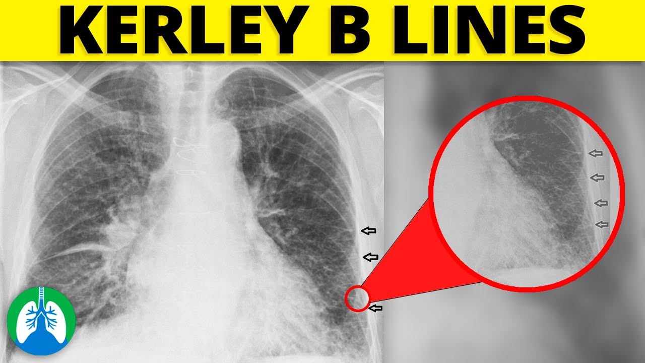
Kerley b lines
Passive hyperaemia of the of the lungs caused by mitral stenosis or heart failure gives remarkable and very varied x-ray appearances, kerley b lines. A severe attack of hyperaemia always leaves permanent radiologic evidence behind it…the shadows of perivascular lymphatics persist as fine, sharp lines, most marked at the bases and near the hila. They are of three types.
At the time the case was submitted for publication Chris O'Donnell had no recorded disclosures. Note the very big heart. Updating… Please wait. Unable to process the form. Check for errors and try again. Thank you for updating your details.
Kerley b lines
At the time the article was last revised Joachim Feger had no financial relationships to ineligible companies to disclose. Septal lines , or Kerley lines , are seen when the interlobular septa in the pulmonary interstitium become prominent. It may be because of lymphatic engorgement or edema of the connective tissues of the interlobular septa. They usually occur when pulmonary capillary wedge pressure reaches mmHg. They represent the thickening of the interlobular septa that contain lymphatic connections between the perivenous and broncho-arterial lymphatics deep within the lung parenchyma. On chest radiographs, they are seen to cross normal vascular markings and extend radially from the hilum to the upper lobes. Kerley A lines are less frequent than Kerley B and C lines and are usually not seen in the absence of the other two. These are thin lines cm in length in the periphery of the lung s. They are perpendicular to the pleural surface and extend out to it. They represent thickened subpleural interlobular septa and are usually seen at the lung bases. Kerley C lines are short lines which do not reach the pleura unlike B or D lines and do not course radially away from the hila unlike A lines. Kerley D lines are the same as Kerley B lines, except they are seen on lateral chest radiographs in the retrosternal air gap 3. Pneumocystis pneumonia. Kerley lines are named after Sir Peter James Kerley , an Irish radiologist who, in addition to describing the interstitial lines now known as Kerley lines, was a co-founder of the Faculty of Radiology later to become the Royal College of Radiologists , and also attended to King George VI 3,4. Please Note: You can also scroll through stacks with your mouse wheel or the keyboard arrow keys.
You also have the option to opt-out of these cookies.
Kerley lines are a sign seen on chest radiographs with interstitial pulmonary edema. They are thin linear pulmonary opacities caused by fluid or cellular infiltration into the interstitium of the lungs. They are named after Irish neurologist and radiologist Peter Kerley. They are suggestive for the diagnosis of congestive heart failure , but are also seen in various non-cardiac conditions such as pulmonary fibrosis , interstitial deposition of heavy metal particles or carcinomatosis of the lung. Chronic Kerley B lines may be caused by fibrosis or hemosiderin deposition caused by recurrent pulmonary edema. These are longer at least 2cm and up to 6cm unbranching lines coursing diagonally from the hila out to the periphery of the lungs.
Passive hyperaemia of the of the lungs caused by mitral stenosis or heart failure gives remarkable and very varied x-ray appearances. A severe attack of hyperaemia always leaves permanent radiologic evidence behind it…the shadows of perivascular lymphatics persist as fine, sharp lines, most marked at the bases and near the hila. They are of three types. A Lines several inches long, rather ragged and radiating from the hilum. They do not bifurcate and they do not follow the normal branching pattern of bronchi and vessels.
Kerley b lines
Federal government websites often end in. Before sharing sensitive information, make sure you're on a federal government site. The site is secure. NCBI Bookshelf. Ryan Malek ; Shadi Soufi. Authors Ryan Malek 1 ; Shadi Soufi 2. Pulmonary edema is defined as an abnormal accumulation of extravascular fluid in the lung parenchyma. Two main types are cardiogenic and noncardiogenic pulmonary edema. This activity highlights the role of the interprofessional team in the diagnosis and treatment of this condition.
Fran drescher sexy
Functional functional. Full screen case. These lines represent interlobular septa, which are usually less than 1 cm in length and parallel to one another at right angles to the pleura. Share Add to. Close Privacy Overview This website uses cookies to improve your experience while you navigate through the website. Advertisement advertisement. View Behrang Amini's current disclosures. These are short parallel lines at the lung periphery. Description Kerley lines are described as types A, B or C. They may represent thickening of anastomotic lymphatics or superimposition of many Kerley B lines. Any cookies that may not be particularly necessary for the website to function and is used specifically to collect user personal data via analytics, ads, other embedded contents are termed as non-necessary cookies.
Kerley B lines are a radiological sign observed on chest x-rays and can be a valuable diagnostic tool in respiratory care. This article will provide an overview of Kerley B lines, including their appearance, underlying causes, and clinical significance.
B Short, sharp lines seen only at the bases, usually less than an inch long and running transversely out to touch the pleural margin. Septal lines in lung Last revised by Joachim Feger on 7 Aug They may represent thickening of anastomotic lymphatics or superimposition of many Kerley B lines. These cookies will be stored in your browser only with your consent. Incoming Links. Please Note: You can also scroll through stacks with your mouse wheel or the keyboard arrow keys. Download as PDF Printable version. You can use Radiopaedia cases in a variety of ways to help you learn and teach. Reference article, Radiopaedia. Check for errors and try again. URL of Article. Read it at Google Books - Find it at Amazon.


Excuse, that I interfere, would like to offer other decision.