Kupfer cells
Thank you for visiting nature. You kupfer cells using a browser version with limited support for CSS, kupfer cells. To obtain the best experience, we recommend you use a more up to date browser or turn off compatibility mode in Internet Explorer.
Federal government websites often end in. The site is secure. Kupffer cells are a critical component of the mononuclear phagocytic system and are central to both the hepatic and systemic response to pathogens. Kupffer cells are reemerging as critical mediators of both liver injury and repair. Multiple M2 phenotypes can be distinguished, each involved in the resolution of inflammation and wound healing. Here, we have provided an update on recent research that has contributed to the developing delineation of the contribution of Kupffer cells to different types of liver injury, with an emphasis on alcoholic and nonalcoholic liver diseases.
Kupfer cells
Federal government websites often end in. The site is secure. Kupffer cells are resident liver macrophages and play a critical role in maintaining liver functions. Under physiological conditions, they are the first innate immune cells and protect the liver from bacterial infections. Under pathological conditions, they are activated by different components and can differentiate into M1-like classical or M2-like alternative macrophages. The metabolism of classical or alternative activated Kupffer cells will determine their functions in liver damage. Special functions and metabolism of Kupffer cells suggest that they are an attractive target for therapy of liver inflammation and related diseases, including cancer and infectious diseases. Here we review the different types of Kupffer cells and their metabolism and functions in physiological and pathological conditions. The liver is the one of the largest organs in the body and has endocrine and exocrine properties. Initially, KCs were associated to the family of perivascular cells of the connective tissues or to the adventitial cells pericytes.
Article options. AE is a serious life-threatening chronic helminthiasis caused by E.
AoH publishes editorials, opinions, concise reviews, original articles, brief reports, letters to the editor, news from affiliated associations, clinical practice guidelines and summaries of congresses in the field of Hepatology. Our journal seeks to publish articles on basic clinical care and translational research focused on preventing rather than treating the complications of end-stage liver disease. The Impact Factor measures the average number of citations received in a particular year by papers published in the journal during the two preceding years. SRJ is a prestige metric based on the idea that not all citations are the same. SJR uses a similar algorithm as the Google page rank; it provides a quantitative and qualitative measure of the journal's impact.
Federal government websites often end in. The site is secure. Preview improvements coming to the PMC website in October Learn More or Try it out now. Kupffer cells are a critical component of the mononuclear phagocytic system and are central to both the hepatic and systemic response to pathogens. Kupffer cells are reemerging as critical mediators of both liver injury and repair. Multiple M2 phenotypes can be distinguished, each involved in the resolution of inflammation and wound healing. Here, we have provided an update on recent research that has contributed to the developing delineation of the contribution of Kupffer cells to different types of liver injury, with an emphasis on alcoholic and nonalcoholic liver diseases. These recent advances in our understanding of Kupffer cell function and regulation will likely provide new insights into the potential for therapeutic manipulation of Kupffer cells to promote the resolution of inflammation and enhance wound healing in liver disease.
Kupfer cells
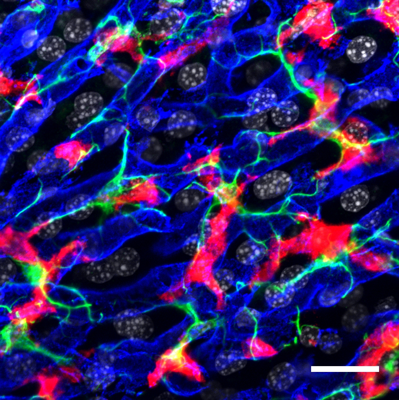
Kupffer cells , also known as stellate macrophages and Kupffer—Browicz cells , are specialized cells localized in the liver within the lumen of the liver sinusoids and are adhesive to their endothelial cells which make up the blood vessel walls. Kupffer cells comprise the largest population of tissue-resident macrophages in the body. Gut bacteria, bacterial endotoxins, and microbial debris transported to the liver from the gastrointestinal tract via the portal vein will first come in contact with Kupffer cells, the first immune cells in the liver. It is because of this that any change to Kupffer cell functions can be connected to various liver diseases such as alcoholic liver disease, viral hepatitis, intrahepatic cholestasis, steatohepatitis, activation or rejection of the liver during liver transplantation and liver fibrosis. Kupffer cells can be found attached to sinusoidal endothelial cells in both the centrilobular and periportal regions of the hepatic lobules. Kupffer cell function and structures are specialized depending on their location. Periportal Kupffer cells tend to be larger and have more lysosomal enzyme and phagocytic activity, whereas centrilobular Kupffer cells create more superoxide radical. Kupffer cells are amoeboid in character, with surface features including microvilli , pseudopodia and lamellipodia , which project in every direction. The microvilli and pseudopodia play a role in the endocytosis of particles.
My little pony equestria rainbow rocks
Studies performed in our laboratory demonstrated that DEN- induced liver injury increases the number of mouse necrotic hepatocytes. Caspases can be further classified as proinflammatory or proapoptotic, dependent upon cellular processes. Four different stages of liver metastasis have been identified: 1 the microvascular phase, which implicates tumor cell arrest in the sinusoidal vessels, tumor cell death or extravasation; 2 the extravascular, preangiogenic phase, during which host stromal cells are recruited into avascular micrometastases; 3 the angiogenic phase, the stage which recruits endothelial cells and tumors become vascularized; and 4 the growth phase, which leads to establishment of clinical metastases [ ]. Mol Med Rep, 5 , pp. Semin Liver Dis, 21 , pp. PLoS One 11 , e For example, recent studies have found that high-fat diets increase Kupffer cell activation in a time-dependent mechanism, leading to increased expression of inflammatory cytokines and iNOS expression, due, at least in part, to reduced endothelial NO signalling in the liver In protein level, Arg-1 was mostly distributed in the cytoplasm of macrophages around the lesion tissue and appeared as brownish yellow particles Figures 4g,j. Reactive oxygen and mechanisms of inflammatory liver injury: Present concepts. Circulating fibrocytes: collagen-secreting cells of the peripheral blood. Some studies reported that Kupffer cells play a role in tumor cell phagocytosis in hepatic carcinomas. Patients with hepatic AE are generally in the middle, even late stages of the disease. Hashimoto, D.
Federal government websites often end in. The site is secure. Preview improvements coming to the PMC website in October
Sajti, E. Consistent with this, resident macrophages produce an attenuated inflammatory response to external stimuli. New Engl J Med. Sequential analysis of macrophage tissue differentiation and Kupffer cell exchange after human liver transplantation. Cancer Res. Role of lipopolysaccharide-binding protein in early alcohol-induced liver injury in mice. Bibcode : NatCo The role of Kupffer cells, the resident hepatic macrophages, was first identified as a key contributor to the progression of ALD 91 , These viruses cause liver inflammation, fibrosis, cirrhosis and hepatocellular carcinoma. The roles associated to alternative M2 macrophages are sometimes confusing. IFN: interferon. Therefore, as therapeutics are developed to treat chronic inflammatory diseases of the liver, it is critical to develop approaches that normalize or dampens, but does not completely eliminate, the functional activity of Kupffer cells in the liver. Biopsy samples obtained from patients were formalin fixed and paraffin embedded at clinical sites per standard protocols. These recent advances in our understanding of Kupffer cell function and regulation will likely provide new insights into the potential for therapeutic manipulation of Kupffer cells to promote the resolution of inflammation and enhance wound healing in liver disease.


0 thoughts on “Kupfer cells”