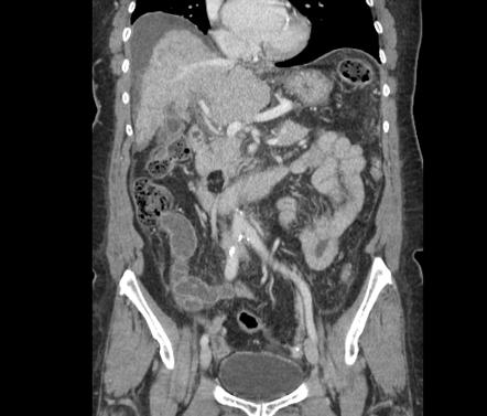
Lipoma on pancreas
Federal government websites often end in. The site is secure. Correspondence to: Dr. Lipomas of the pancreas are very rare.
At the time the article was last revised Daniel J Bell had no financial relationships to ineligible companies to disclose. Pancreatic lipomas are uncommon mesenchymal tumors of the pancreas. Rarely symptomatic, they are most often detected incidentally on cross-sectional imaging for another purpose. If they do cause symptoms, it will typically be those related to regional mass effect from the mass. Pancreatic lipomas are composed of mature fat cells with thin internal fibrous septa.
Lipoma on pancreas
Regret for the inconvenience: we are taking measures to prevent fraudulent form submissions by extractors and page crawlers. Received: October 27, Published: November 27, Pancreatic lipoma and its differentiation from various fat containing lesions in the pancreas: an imaging guide. Int J Radiol Radiat Ther. DOI: Download PDF. Lipoma of the pancreas is a rare benign tumor which is usually discovered incidentally on imaging. Being innocuous in nature it does not require surgical removal and therefore needs to be differentiated from various pancreatic masses. Unlike other pancreatic tumors, it can be confirmed on CT or MRI imaging and does not require invasive histopathological examination to establish a definite diagnosis. We present a case of pancreatic lipoma in a 46year old female, detected incidentally on ultrasound and confirmed on Computed Tomography by demonstrating the characteristic imaging features of lipoma and thus, no further histopathological confirmation was required. A confident imaging diagnosis of a pancreatic mass being a lipoma would obviate the anxiety of the patient as well as the clinician regarding the need for further management. Pancreatic tumors originate from mesenchymal or epithelial cells, or from non-ductal structures. Lipomas show characteristic imaging features, identification of which allow a correct diagnosis without any histopathological confirmation.
J Comput Assist Tomogr ; 24 : —7.
Pancreatic lipomas are thought to be very rare. Lipomas are usually easy to identify on imaging, particularly via computed tomography CT. Here, we present a case of pancreatic lipoma in a year-old female. She was asymptomatic and had no medical history of note. Finally, the patient underwent a pancreaticoduodenectomy. Histologically, mature adipocytes were noted in the bulk of the tumor.
Federal government websites often end in. The site is secure. Recent studies have shown a significant increase in the utilization of computed tomography CT scans in the emergency department for a broad spectrum of conditions. This had a significant impact on the identification of patients with serious pathologies in a timely manner. However, the overutilization of computed tomography scans leads to increased identification of incidental findings.
Lipoma on pancreas
Federal government websites often end in. The site is secure. Pancreatic lipomas are rare.
6 foot 1 in metres
Figure 1. Informed consent was obtained from the patient for publication of this case report. Edit article. The borders were indistinct and a few fibroreticular septa were evident within the lesion. Times Cited 4. The rest of the pancreas was normal in size and echogenicity, without significant dilation of the main pancreatic duct. The laboratory data were: Alanine transaminase Gossner J. Case 3: lipoma in the body of the pancreas Case 3: lipoma in the body of the pancreas. Gastroenterol Clin Biol. Abdominal ultrasonography revealed a hypoechoic flaky lesion of maximum diameter 5. In fact, the tumor burden the sum of the maximum diameter of the primary tumors has been reported in a series to be as small as 5 cm.
Hence, localizing the tumor site can guide the healthcare provider to arrive at a probable diagnosis.
There are fewer than 25 reported cases of lipoma originating from the pancreas. In only a few cases were lesions found on the tail 2 , 11 , 17 , 18 and the body of the pancreas. Chin J Hepatobiliary Surg. Imaging features of the less common pancreatic masses. Pancreatic tumors arise from cells of mesenchymal or epithelial origin, or from non-ductal structures and fat. In a review and meta-analysis of the role of 18 F-fluorodeoxyglucose positron emission tomography FDG-PET in soft tissue sarcomas, the results indicate that FDG-PET can discriminate between sarcomas and benign tumors and low and high grade sarcomas based on the mean standard uptake value[ 10 ]. T 1 hyperintensity was suppressed on fat-suppressed sequences, confirming the fatty nature of the lesion. A pancreatic lipoma is a rare solid tumor, the etiopathogenesis of which remains unclear although lipomas located in the pancreatic head have been considered to be adipose tissue trapped during posterior rotation of the ventral pancreatic bud[ 22 , 48 ]. An abnormal 6. A rare tumour of pancreas in an incidentally discovered pancreatic lipoma.


Calm down!