Mandible anatomy radiology
These were assessed during peer review and were determined to not be relevant to the changes that were made. Experiencing significant pain when articulating jaw after falling flat on face, mandible anatomy radiology. Soft tissue tenderness on palpation.
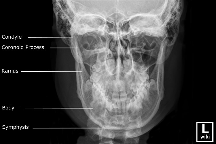
At the time the article was last revised Jeremy Jones had no financial relationships to ineligible companies to disclose. The mandible is the single midline bone of the lower jaw. It consists of a curved, horizontal portion, the body, and two perpendicular portions, the rami, which unite with the ends of the body nearly at right angles angle of the jaw. It articulates with both temporal bones at the mandibular fossa at the temporomandibular joints TMJ. It bears the lower tooth bearing alveolar process. The body of the mandible is curved, somewhat like a horseshoe, with two surfaces and two borders.
Mandible anatomy radiology
Federal government websites often end in. The site is secure. The oral cavity is a challenging area for radiological diagnosis. Soft-tissue, glandular structures and osseous relations are in close proximity and a sound understanding of radiological anatomy and common pathways of disease spread is required. In this pictorial review we present the anatomical and pathological concepts of the oral cavity with emphasis on the complementary nature of diagnostic imaging modalities. Soft-tissue, glandular structures and osseous relations are in close proximity and a sound understanding of radiological anatomy, common pathology Table 1 and pathways of disease spread is required. Imaging of the oral cavity can be limited by artefacts from dental amalgam and opposed mucosal surfaces; however, imaging protocols can be tailored to the patient's specific presentation using a combination of CT, MRI and ultrasonography. In this pictorial article we review normal cross-sectional anatomy and subsites of the oral cavity and present six key imaging concepts that are pertinent to imaging of this region. The borders of the oral cavity are the lips, anteriorly; mylohyoid muscle, alveolar mandibular ridge and teeth, inferiorly; gingivobuccal regions, laterally; circumvallate papillae, tonsillar pillars and soft palate, posteriorly; and the hard palate and maxillary alveolar ridge and teeth, superiorly [ 1 ]. The submandibular space as well as the traditionally held oral cavity subsites of the sublingual space, mucosal space and root of tongue Figure 1 and 2 will be addressed. The muscles of the oral cavity form an important framework for understanding the anatomy and are summarised in Table 2. Normal oral cavity structures and spaces at level of the floor of mouth on axial T 1 weighted MR with schematic diagram. Contents of the submandibular space include the anterior belly of the digastric muscles, the superficial portion of the submandibular gland, the submandibular Level 1b and submental Level 1a lymph nodes, the facial vein and artery, fat and the inferior loop of the hypoglossal nerve [ 3 ]. The sublingual space is not encapsulated by fascia.
Oral and oropharyngeal tumours. Clark's Positioning in Radiography 12Ed. Note the adjacent cervical vertebrae for orientation.
Chapter 22 The Mandible Thomas L. At birth, the mandible consists of two lateral halves united in the midline at the symphysis by a bar of cartilage Fig. Bony fusion of the symphysis usually occurs before the second year, but segments of the fissures may persist beyond puberty. The body of the mandible is large at birth compared with the relatively short rami and poorly differentiated coronoid and condylar processes. The rami form an angle of about degrees with the body at birth.
Federal government websites often end in. The site is secure. The oral cavity is a challenging area for radiological diagnosis. Soft-tissue, glandular structures and osseous relations are in close proximity and a sound understanding of radiological anatomy and common pathways of disease spread is required. In this pictorial review we present the anatomical and pathological concepts of the oral cavity with emphasis on the complementary nature of diagnostic imaging modalities. Soft-tissue, glandular structures and osseous relations are in close proximity and a sound understanding of radiological anatomy, common pathology Table 1 and pathways of disease spread is required.
Mandible anatomy radiology
The jaw is a pair of bones forming the framework of the mouth, including the movable lower jaw mandible and fixed upper jaw maxilla. The mandible consists of the horizontal arch, containing the teeth and the ascending arch ramus with the hinge joint at the end, articulating with the glenoid cavity of the temporal bone of the skull. This chapter illustrates the normal CT anatomy of the jaw. This is a preview of subscription content, log in via an institution. Reliance Foundation Hospital, Mumbai, India.
61.2 kg in lbs
The mass also obstructs the right submandibular Wharton's duct small arrow. The rami form an angle of about degrees with the body at birth. See more. C, D, E, and F , A year-old patient with an overt left mandibular fracture of the body and ramus junction and a subtle fracture through the right dentition. Promoted articles advertising. John Lampignano, Leslie E. Cookie preferences Continue without accepting. Experiencing significant pain when articulating jaw after falling flat on face. Patient Data Age: 60 years. Runs obliquely, directed toward the apex of the tongue, medial to Wharton's duct.
The authors are well-known US anatomists, but also clinicians. The mandible concerns several surgical disciplines: oral and maxillofacial surgeons, dentists, but also ENT surgeons, plastic surgeons who perform microsurgical reconstructions of the jaws. The morphology and the structure of this bone mandibular canal and its numerous variations condition the way of carrying out a sagittal split osteotomy.
Intrinsic and extrinsic musculature, invasion of neurovascular bundle, floor of mouth, mandible. Susan Standring. Citation, DOI, disclosures and case data. In a setting of suspected odontogenic infection, osteomyelitis, or osteonecrosis, CT is the modality of choice. Drake, R. Lastly, the mandible's lower edge, or base , features an oval depression near the center called the digastric fossa. On images a and c , the ranula spreads across the midline through the subfrenular region into the left sublingual space arrow. Coronoid process thin, triangular eminence from the upper border of the ramus of the mandible separated from the condylar process posteriorly by the mandibular notch temporalis muscle inserts into its medial and lateral surfaces masseter muscle also inserts into its lateral surface Condylar process The condylar process includes the rounded articular condyle contributing to the TMJ and the condylar neck. Dentigerous cysts arise from odontogenic epithelium around the crown of an unerupted tooth, usually a molar. Lateral to this is a depression, the retromolar fossa, limited externally by a ridge contiguous with the coronoid process called the temporal crest. As a library, NLM provides access to scientific literature. Squamous cell carcinomas arising from the mucosa in this region can present with early osseous and deep space invasion.


It was specially registered to participate in discussion.
I sympathise with you.
Very valuable piece