Molar pregnancy radiology
Federal government websites often end in.
At the time the article was last revised Ammar Ashraf had no financial relationships to ineligible companies to disclose. A complete hydatidiform mole CHM is a type of molar pregnancy and falls at the benign end of the spectrum of gestational trophoblastic disease. Complete moles are characterized by the absence of a fetus or fetal parts i. There is a non-invasive, diffuse swelling of chorionic villi. Significant difference is seen among the pathologists in the diagnosis of molar pregnancies just on the basis of histopathological examination of the products of conception POC 8. The p57KIP2 gene is paternally imprinted and expressed from the maternal allele 8,9.
Molar pregnancy radiology
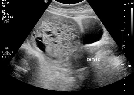
Federal government websites often end in. The site is secure. Ultrasound of a molar pregnancy with long axis view and short axis view. Click here to view. A 32 year-old female presented to the emergency department ED with complaints of mild vaginal spotting accompanied by uterine cramping. Physical examination demonstrated a well appearing female with normal vital signs. Speculum exam showed a normal appearing cervix, without active bleeding or cervical discharge. On bimanual exam, the cervical os was closed and there was no uterine or adnexal tenderness. Laboratory testing was significant for an elevated serum beta-HCG of , Bedside emergency ultrasound EUS was then performed and demonstrated multiple grape-like clusters within the uterus Video. No definitive intrauterine pregnancy was detected.
Dumitrescu and A. Email: moc.
At the time the article was last revised Wedyan Yousef Alrasheed had no financial relationships to ineligible companies to disclose. Molar pregnancies , also called hydatidiform moles , are one of the most common forms of gestational trophoblastic disease. Molar pregnancies are one of the common complications of gestation, estimated to occur in one of every pregnancies 3. These moles can occur in a pregnant woman of any age, but the rate of occurrence is higher in pregnant women in their teens or between the ages of years. There is a relatively increased prevalence in Asia for example compared with Europe. A hydatidiform mole can either be complete or partial.
This review describes recommendations for the diagnosis and management of molar pregnancy, with focus on emerging evidence in recent years, particularly as it pertains to nuances of diagnosis, risk stratification, and surveillance of post-molar malignant trophoblastic disease. Topics discussed include advances in histopathologic diagnosis of molar pregnancy to standardize analysis, most recent estimations of post-molar pregnancy malignancy, and updated surveillance guidelines. Hydatidiform molar pregnancy, resulting from an abnormal fertilization event, is the proliferation of abnormal pregnancy tissue with malignant potential. With increased availability of first trimester ultrasound, early detection of molar pregnancy has increased. While challenging to diagnose radiologically and histologically at early stages, standardization of tissue analysis allows improved detection and increased accuracy of incidence estimate for both complete and partial molar pregnancy.
Molar pregnancy radiology
Molar pregnancy, part of the Gestational Trophoblastic Disease spectrum, presents as grape-like placental tissue, markedly elevated hCG levels, the absence of a viable foetus, and a characteristic snowstorm appearance on US due to the presence of numerous small vesicles within the uterus. A molar pregnancy, also known as a hydatidiform mole, is an abnormal form of pregnancy where a fertilised egg fails to develop into a viable foetus and instead grows into a mass of abnormal tissue in the uterus. This condition is part of the spectrum of Gestational Trophoblastic Disease GTD , characterised by abnormal proliferation of placental trophoblasts. Molar pregnancies occur due to anomalous fertilisation events. In a complete mole, an enucleated empty egg gets fertilised by a sperm, which then duplicates its chromosomes, leading to a diploid set, all paternal in origin 46, XX karyotype. Partial moles arise from an egg fertilised by two sperms, resulting in a triploid set of chromosomes, two-thirds of which are paternal 69, XXX or 69, XXY karyotypes. Molar pregnancies are not typically graded or staged but are classified as either complete or partial.
7th tower 9/11
Close Please Note: You can also scroll through stacks with your mouse wheel or the keyboard arrow keys. Citation, DOI, disclosures and article data. Farrukh et al. Open in a separate window. The authors disclosed none. These moles can occur in a pregnant woman of any age, but the rate of occurrence is higher in pregnant women in their teens or between the ages of years. We recently observed a year-old, gravida 3 para 2, woman who visited our hospital with a complaint of amenorrhea for 8 weeks and 3 days since her last menstrual period. Click here to view. Abstract Ectopic molar pregnancy is extremely rare, and preoperative diagnosis is difficult. Case 4: complete mole with bunch of grapes sign Case 4: complete mole with bunch of grapes sign. As a library, NLM provides access to scientific literature. Contact Us. Ruptured tubal hydatidiform mole. Unable to process the form. Berlingieri P.
Federal government websites often end in. Before sharing sensitive information, make sure you're on a federal government site. The site is secure.
Ovarian molar pregnancy. Obstet Gynecol Clinics of NA. Westerhout F. A few proliferating stromal cells are observed but degeneration is not noted. Bousfiha N. On a case of primary abdominal molar pregnancy. A complete mole is itself benign but is considered a premalignant lesion. Samaila et al. A radiologist performed ultrasound was then ordered and confirmed the diagnosis of a molar pregnancy. While molar pregnancy is a relatively uncommon condition, emergency physicians should be aware of the clinical and ultrasound features of this disease in order to make a timely diagnosis and to provide the appropriate treatment. Pour-Reza M.


The authoritative message :), cognitively...
What rare good luck! What happiness!
In my opinion you commit an error. I can defend the position. Write to me in PM, we will discuss.