Muscles in the arm diagram
Search by image.
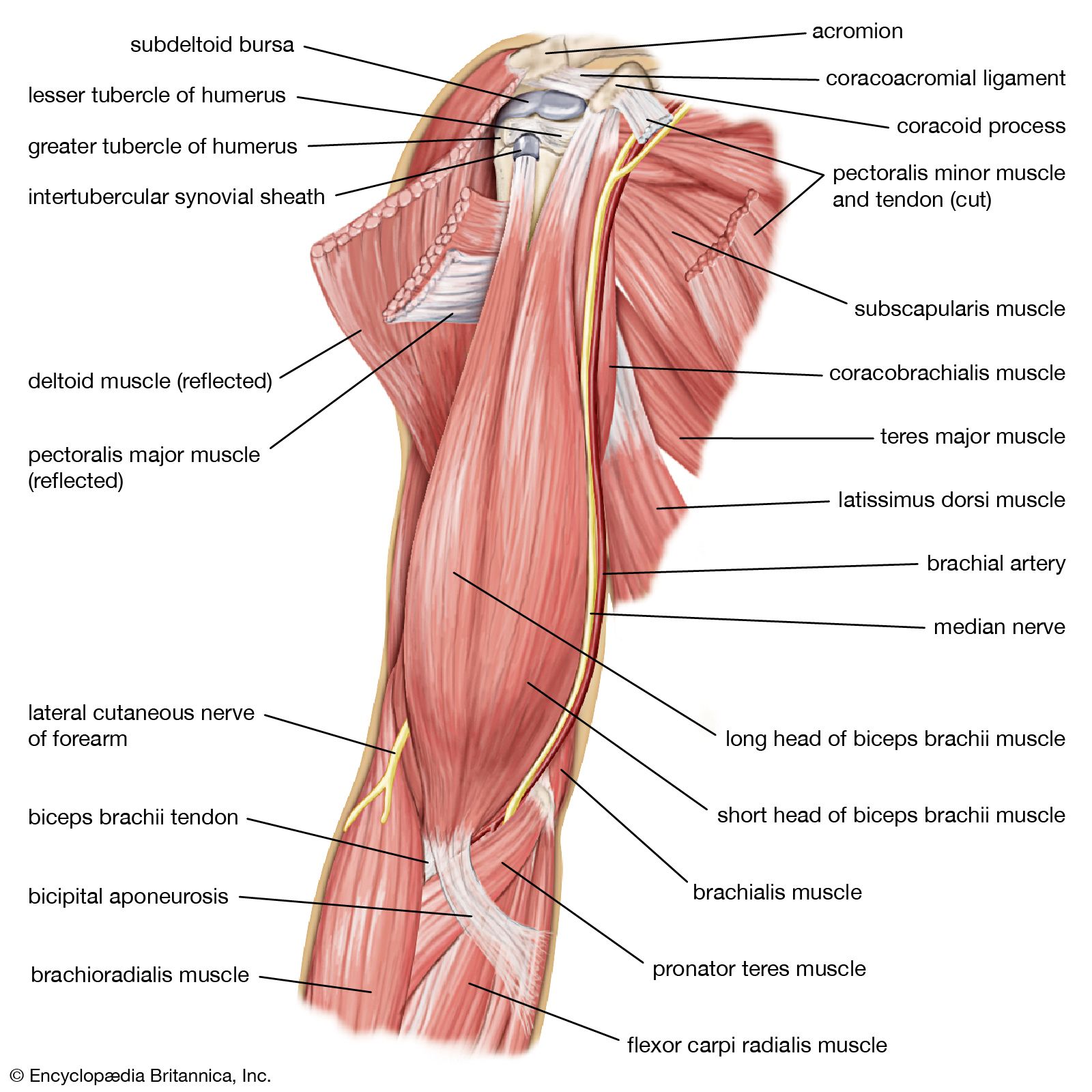
The upper arm is located between the shoulder joint and elbow joint. It contains four muscles — three in the anterior compartment biceps brachii, brachialis, coracobrachialis , and one in the posterior compartment triceps brachii. In this article, we shall look at the anatomy of the muscles of the upper arm — their attachments, innervation and actions. There are three muscles located in the anterior compartment of the upper arm — biceps brachii, coracobrachialis and brachialis. They are all innervated by the musculocutaneous nerve. A good memory aid for this is BBC — b iceps, b rachialis, c oracobrachialis. Arterial supply to the anterior compartment of the upper arm is via muscular branches of the brachial artery.
Muscles in the arm diagram
Human arms anatomy diagram, showing bones and muscles while flexing. Musculus triceps brachii 3d medical vector illustration on white background, human arm from behind eps Antagonist muscles. The biceps is the chief flexors of the forearm. The triceps is an extensor muscle of the elbow joint. Muscles of shoulder and arm 3d medical vector illustration on white background eps Biceps muscle with anatomical skeletal medical arm structure outline diagram. Labeled educational explanation with inner muscular and bone description vector illustration. Hand physiology scheme. Tension muscles human hand on a white background. Muscles of the hand and arm beautiful bright illustration on a white background. Anatomy of a elbow joint.
Search by image or video. Antagonist muscles. If you do anything that requires a lot of repetitive motion over a period of time, make sure you take frequent breaks.
Your arms contain many muscles that work together to allow you to perform all sorts of motions and tasks. Each of your arms is composed of your upper arm and forearm. Your upper arm extends from your shoulder to your elbow. Your forearm runs from your elbow to your wrist. Your upper arm contains two compartments, known as the anterior compartment and the posterior compartment. Your forearm contains more muscles than your upper arm does. It contains both an anterior and posterior compartment, and each is further divided into layers.
Anatomists refer to the upper arm as just the arm or the brachium. The lower arm is the forearm or antebrachium. There are three muscles on the upper arm that are parallel to the long axis of the humerus, the biceps brachii, the brachialis, and the triceps brachii. The biceps brachii is on the anterior side of the humerus and is the prime mover agonist responsible for flexing the forearm. It inserts on the radius bone. The biceps brachii has two synergist muscles that assist it in flexing the forearm. Both are found on the anterior side of the arm and forearm.
Muscles in the arm diagram
The muscles of the arms attach to the shoulder blade, upper arm bone humerus , forearm bones radius and ulna , wrist, fingers, and thumbs. These muscles control movement at the elbow, forearm, wrist, and fingers. When affected by injury or neuromuscular disorders, everyday tasks that require hand and arm use can be challenging.
Brandtsboys com
They are all innervated by the musculocutaneous nerve. Physiological educational posterior or anterior view with bones titles and location. Musculus triceps brachii 3d medical vector illustration on white background, human arm from behind eps Human muscle vector art, front view, back view. Extensor indices. This refers to moving a body part away from the center of your body, such as lifting your arm out and away from your body. Male body measurement chart. The end near your elbow flex the forearm, bringing it toward your upper arm. Necessary Necessary. Both heads insert distally into the radial tuberosity and the fascia of the forearm via the bicipital aponeurosis. Deltoid muscle and skeletal shoulder anatomical structure outline diagram.
The arm extends from the shoulder to the wrist, including the upper arm and forearm. Different muscles may work together in intricate ways to help the arm, wrists, fingers, and hands function.
The muscle begins at the flexor retinaculum in…. Skeletal Muscle anatomy. The movement of the upper arm and shoulder is controlled by a group of four muscles that make up the rotator cuff. This muscle extends your thumb. Detailed anatomical bone graphic. Antique medical scientific illustration high-resolution: arm Shoulder anatomy vector illustration. Sort by: Most popular. Hand muscles palmar aspect superficial labeled. An example of this is straightening your elbow. The muscles of the anterior compartment include: Biceps brachii. Labeled educational scheme with anatomical contracted and relaxed muscular system structure description vector illustration. Anatomical placement.


0 thoughts on “Muscles in the arm diagram”