Neuropathic joint radiology
The radiographic features of a Charcot joint can be remembered by using the following mnemonics :. Articles: Charcot joint causes mnemonic Charcot joint Cases: Charcot foot Milwaukee shoulder Charcot foot Diabetic foot Charcot joint - foot Spinal dysraphism with neuropathic bladder and charcot joint Charcot joint - foot Neuropathic Charcot arthopathy of spine, neuropathic joint radiology, knee and neuropathic joint radiology Charcot joint ankle Charcot joint Bilateral Charcot joints Multiple choice questions: Question Please Note: You can also scroll through stacks with your mouse wheel or the keyboard arrow keys. Updating… Please wait.
At the time the article was last revised Mohammadtaghi Niknejad had no financial relationships to ineligible companies to disclose. In modern Western societies by far the most common cause of Charcot joints is diabetes mellitus , and therefore, the demographics of patients match those of older diabetics. Prevalence differs depending on the severity of diabetes mellitus 1 :. Patients present insidiously or are identified incidentally, or as a result of investigation for deformities. Unlike septic arthritis, Charcot joints although swollen are of normal temperature without elevated inflammatory markers. Importantly, they are painless.
Neuropathic joint radiology
Federal government websites often end in. The site is secure. Charcot foot pied de Charcot CF , first described by Jean-Martin Charcot in , is caused by a wide variety of disorders that ultimately destroy the protective mechanisms of the small joints of the foot. Leprosy and diabetes are the most common causes of this form of destructive neuroarthropathy in the developing world. If the diagnosis is missed early in the natural course of the disease, severe foot deformity and disability, ulceration, infection, and ultimately limb amputation are the expected outcomes. Five distinct patterns of involvement have been described in people with diabetes presenting with CF 2. In this article, we share clinical and radiological photographs of each of these subtypes through five case presentations of patients with longstanding diabetes and clinical evidence of advanced peripheral neuropathy in the absence of peripheral vascular disease. A year-old man with type 2 diabetes presented with swelling of the left great toe and a discharging, nonhealing ulcer on its plantar aspect Figure 1. Clinical and radiological examinations were suggestive of osteomyelitis of the left great toe. However, we also noticed mid- and forefoot widening on the right side. Case 1. A Swelling of left great toe with discharging wound black solid arrow and right-sided fore-foot widening white solid arrow. A year-old man with a year history of type 2 diabetes presented with progressive left foot deformity for 6 months after a trivial trauma and subsequent development of a nonhealing ulcer over the midfoot for the past 3 months.
Information collected from Ahmadi et al.
Federal government websites often end in. The site is secure. Data sharing is not applicable to this article as no datasets were generated or analyzed. Charcot foot refers to an inflammatory pedal disease based on polyneuropathy; the detailed pathomechanism of the disease is still unclear. Patients with Charcot foot typically present in their fifties or sixties and most of them have had diabetes mellitus for at least 10 years. If left untreated, the disease leads to massive foot deformation. This review discusses the typical course of Charcot foot disease including radiographic and MR imaging findings for diagnosis, treatment, and detection of complications.
Are you sure you want to trigger topic in your Anconeus AI algorithm? Would you like to start learning session with this topic items scheduled for future? Please confirm topic selection. No Yes. Please confirm action. You are done for today with this topic. Cards Cards. Questions Questions. Cases Cases.
Neuropathic joint radiology
At the time the article was last revised Mohammadtaghi Niknejad had no financial relationships to ineligible companies to disclose. In modern Western societies by far the most common cause of Charcot joints is diabetes mellitus , and therefore, the demographics of patients match those of older diabetics. Prevalence differs depending on the severity of diabetes mellitus 1 :. Patients present insidiously or are identified incidentally, or as a result of investigation for deformities. Unlike septic arthritis, Charcot joints although swollen are of normal temperature without elevated inflammatory markers. Importantly, they are painless. The pathogenesis of a Charcot joint is thought to be an inflammatory response from a minor injury that results in osteolysis. In the setting of peripheral neuropathy, both the initial insult and inflammatory response are not well appreciated, allowing ongoing inflammation and injury 1. There are two patterns of Charcot joint: atrophic and hypertrophic.
Osrs spottier cape
Download citation. Springer Nature remains neutral with regard to jurisdictional claims in published maps and institutional affiliations. Partha P. It is necessary to use a fluid sensitive sequence e. Cuboid height: perpendicular distance from the plantar aspect of the cuboid to a line drawn from the plantar surface of the calcaneal tuberosity to the plantar aspect of the 5th metatarsal head. Prominent well-marginated subchondral cysts are a typical feature of the chronic Charcot foot Fig. Dynamic contrast enhancement DCE -perfusion may help in the discrimination between viable tissue and necrosis. Pott disease Pott disease. Sanders and Frykberg classification Sanders and Frykberg identified five zones of disease distribution according to their anatomical location, as demonstrated in Fig. In this article, we share clinical and radiological photographs of each of these subtypes through five case presentations of patients with longstanding diabetes and clinical evidence of advanced peripheral neuropathy in the absence of peripheral vascular disease. Copy Download. Last revised:. Culture of the tissue bits obtained from the deeper aspect of the ulcer grew Klebsiella species.
A nonsmoking, man with no previous comorbidities, attended to us for painless inflammation and edema of left ankle and foot for at least 7 months, without fever or other joint swellings. There was no history of trauma.
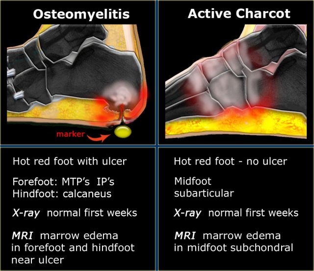
Diagnostic values for skin temperature assessment to detect diabetes-related foot complications. Rocker-bottom deformity: end-stage of Charcot foot. Different thermometers are available to identify subtle differences in surface temperature between two feet and are useful for screening patients in a busy clinic setting. The typical end-stage appearance of a Charcot foot is the so-called rocker-bottom deformity Fig. These fractures are mostly nontraumatic, limited to the posterior third of the bone, and identical to the calcaneal fatigue fracture, with displacement and rotation of the posterior tuberosity. PET-CT allows the quantification of the inflammatory process in all stages of Charcot foot and allows to follow-up its evolution over time: recent research showed that PET-CT may be of additional help for evaluation of treatment duration in addition to MR imaging [ 32 ]. However, there are some imaging features listed in Table 1 , Fig. Metrics details. Bone proliferation and sclerosis, debris, and intraarticular bodies can occur Fig. A year-old man with poorly controlled diabetes presented with a discharging ulcer on the lateral aspect his left hind foot. At the time the article was last revised Mohammadtaghi Niknejad had no financial relationships to ineligible companies to disclose. Natural course of disease Charcot foot is characterized by four different disease stages Fig. Lateral radiograph of the left foot in a patient with Charcot foot involving zone III according to Sanders and Frykberg classification tarsal joints. Patterns I and II are frequently associated with bony deformities and ulcerations, which are considered the cutaneous marker of these forms of CF.


There can be you and are right.
What words... super, an excellent phrase