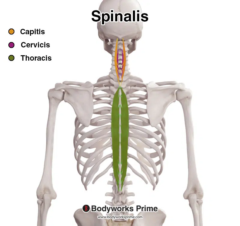
Spinalis origin and insertion
The spinalis is a deep muscle of the back. It is the smallest of the muscle columns within the erector spinae complex, and can be divided into the three parts — thoracic, spinalis origin and insertion, cervicis and capitis although the cervicis part is absent in some individuals.
The spinalis muscle is situated in the middle and upper back as well as the neck, running parallel to the spine. It plays an important role in extending the back and neck, while also aiding in lateral flexion movements. The spinalis muscle is a member of the erector spinae muscle group. The erector spinae muscles consist of the spinalis, iliocostalis , and the longissimus. The erector spinae muscles are deep muscles of the back which run in a vertical direction, parallel to the vertebral column.
Spinalis origin and insertion
The spinalis muscle is the most medial of the erector spinae group of muscles, and is lateral to the multifidus group. The spinalis detaches from medial side of the longissimus thoracis and travels forward near thoracic vertebral spinous processes to cervical vertebral spinous processes. It may be divided into two parts:. In the pig and the horse, the spinalis muscle forms a common muscle belly, therefore sometimes termed as "spinalis thoracic et cervicis thoracic and cervical spinal muscle ", whereas in ruminants and carnivores, the thoracic and cervical spinalis muscles receive additional muscular strands from the the mamillary and transverse processes of some vertebrae, and the fibers of the spinlais musccles are closely related to and often difficult to separate form the semispinalis muscle. Therefore, some authors use the compound name "thoracic and cervical spinal and semispinal muscle" to describe this muscular complex. Origin: extends across the spinous processes of one or more thoracic vertebrae, and sometimes last cervical vertebra. Underlying structures:. IMAIOS and selected third parties, use cookies or similar technologies, in particular for audience measurement. Cookies allow us to analyze and store information such as the characteristics of your device as well as certain personal data e. For more information, see our privacy policy. You can freely give, refuse or withdraw your consent at any time by accessing our cookie settings tool.
Make the changes yourself here! It attaches to the spinous processes of C2, T1-T8 and the occipital bone of the skull. We also use third-party cookies that help us analyze and understand how you use this website.
The spinalis is a portion of the erector spinae , a bundle of muscles and tendons , located nearest to the spine. It is divided into three parts: Spinalis dorsi, spinalis cervicis, and spinalis capitis. Spinalis dorsi, the medial continuation of the sacrospinalis , is scarcely separable as a distinct muscle. It is situated at the medial side of the longissimus dorsi , and is intimately blended with it; it arises by three or four tendons from the spinous processes of the first two lumbar and the last two thoracic vertebrae : these, uniting, form a small muscle which is inserted by separate tendons into the spinous processes of the upper thoracic vertebrae, the number varying from four to eight. It is intimately united with the semispinalis dorsi , situated beneath it. Spinalis cervicis, or spinalis colli, is an inconstant muscle, which arises from the lower part of the nuchal ligament , the spinous process of the seventh cervical, and sometimes from the spinous processes of the first and second thoracic vertebrae , and is inserted into the spinous process of the axis , and occasionally into the spinous processes of the two cervical vertebrae below it. Spinalis capitis biventer cervicis is usually inseparably connected with the semispinalis capitis.
The spinalis is a deep muscle of the back. It is the smallest of the muscle columns within the erector spinae complex, and can be divided into the three parts — thoracic, cervicis and capitis although the cervicis part is absent in some individuals. It is the smallest of the muscle columns within the erector spinae complex, and can be divided into the three parts - thoracic, cervicis and capitis although the cervicis part is absent in some individuals. Once you've finished editing, click 'Submit for Review', and your changes will be reviewed by our team before publishing on the site. We use cookies to improve your experience on our site and to show you relevant advertising. To find out more, read our privacy policy. Spinalis Home Encyclopaedia S Spinalis. Not yet rated. Attachments: Arises from the lower thoracic and lumbar vertebrae, sacrum, posterior aspect of the iliac crest, and the sacroiliac and supraspinous ligament. It attaches to the spinous processes of C2, T1-T8 and the occipital bone of the skull.
Spinalis origin and insertion
Search site Search Search. Go back to previous article. Sign in. Eye Muscle Action Origin Insertion levator palpebrae superioris elevating and retracting the upper eyelid sphenoid bone upper eyelid inferior oblique looking up and laterally eye roll maxilla bone eyeball inferior, lateral inferior rectus looking down depression sphenoid bone eyeball inferior, medial lateral rectus looking laterally abduction sphenoid bone eyeball lateral, anterior medial rectus looking medially adduction sphenoid bone eyeball medial superior oblique looking down and laterally eye roll sphenoid bone eyeball superior, lateral superior rectus looking up elevation sphenoid bone eyeball superior, anterior.
Sorting hat quiz
Found an error? The origins are highlighted in red, while the insertions are colored blue. To find out more, read our privacy policy. Necessary Necessary. Google Analytics. This article incorporates text in the public domain from page of the 20th edition of Gray's Anatomy You can consent to the use of these technologies by clicking "accept all cookies". Read Edit View history. Cookie preferences Continue without accepting. In the pig and the horse, the spinalis muscle forms a common muscle belly, therefore sometimes termed as "spinalis thoracic et cervicis thoracic and cervical spinal muscle ", whereas in ruminants and carnivores, the thoracic and cervical spinalis muscles receive additional muscular strands from the the mamillary and transverse processes of some vertebrae, and the fibers of the spinlais musccles are closely related to and often difficult to separate form the semispinalis muscle. Dorsal branches of the posterior intercostal artery, deep cervical artery, muscular branches of vertebral artery. Anatomical terms of muscle [ edit on Wikidata ]. The content on this website is for informational purposes only and is not intended to be a substitute for professional medical advice, diagnosis, or treatment.
The spinalis Latin: musculus spinalis is one of the muscles forming the erector spinae - a muscle complex consisting of several smaller intrinsic deep back muscle groups that all together form the intermediate layer of the deep back muscles. The other two groups are the longissimus and iliocostalis muscles. The erector spinae muscles run along the length of the spine , and the spinalis is the most medial of the three erector spinae muscles.
The spinalis thoracis section is involved in trunk extension. There are also individual variations between people as outlined below:. Found an error? These cookies will be stored in your browser only with your consent. The spinalis cervicis and capitis sections are involved in neck extension. In Once you've finished editing, click 'Submit for Review', and your changes will be reviewed by our team before publishing on the site. Want some flashcards to help you remember this information? Article Talk. Spinalis cervicis, or spinalis colli, is an inconstant muscle, which arises from the lower part of the nuchal ligament , the spinous process of the seventh cervical, and sometimes from the spinous processes of the first and second thoracic vertebrae , and is inserted into the spinous process of the axis , and occasionally into the spinous processes of the two cervical vertebrae below it. Muscle Fiber Types Explained. The spinalis muscle is situated in the middle and upper back as well as the neck, running parallel to the spine. Note: For the sake of simplicity, only the left side of the muscle is displayed here.


I am sorry, that I interrupt you.