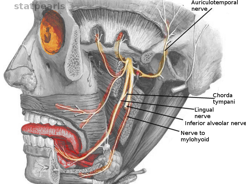
Chorda tympani
The Chorda Tympani Nerve is given off from the facial as it passes downward behind the tympanic cavity, about 6 mm. It then descends between the Pterygoideus externus and internus on the medial surface of the spina chorda tympani of the sphenoid, which it sometimes grooves, and joins, at an acute angle, chorda tympani, the posterior border of the lingual nerve. It receives a few efferent fibers from chorda tympani motor root; these enter the submaxillary ganglion, and through it are distributed to the submaxillary and sublingual glands; the majority of its fibers are afferent, and are continued onward through the muscular substance of the tongue to the mucous membrane covering its anterior two-thirds; they constitute the nerve of taste for this portion of the tongue. Before uniting with the lingual nerve the chorda tympani is joined by a small branch from the otic ganglion, chorda tympani.
Damage can lead to loss of taste, burning mouth syndrome. The chorda tympani is a branch of the facial nerve and, along with other nerves, is important for carrying information about taste and other sensations from your taste buds to your brain. It's also involved in salivary function and a process called inhibition, which means that it lessens signals from other nerves that have to do with both taste and pain. While the cranial nerves themselves are part of the central nervous system, the chorda tympani functions as part of the peripheral nervous system. It's therefore considered a peripheral nerve.
Chorda tympani
These were assessed during peer review and were determined to not be relevant to the changes that were made. The chorda tympani is a nerve that arises from the mastoid segment of the facial nerve , carrying afferent special sensation from the anterior two-thirds of the tongue via the lingual nerve , as well as efferent parasympathetic secretomotor innervation to the submandibular and sublingual glands. After branching off from the facial nerve, the chorda tympani courses through the temporal bone before joining the lingual nerve 2 :. The distance of ascent is variable, depending on the initial branching pattern from the mastoid segment of facial nerve. It then travels inferiorly to join the lingual nerve approximately 2 cm below the skull base. Articles: Middle ear tumours Lingual nerve Anterior tympanic artery Retrotympanum Tongue Parasympathetic nervous system Nervus intermedius Middle ear Mesotympanum Petrotympanic fissure Infratemporal fossa Greater wing of sphenoid Sublingual gland Tympanic membrane Facial nerve Submandibular ganglion Submandibular gland Cases: Anatomy of the genicular ganglion Gray's illustration Trigeminal and facial nerve connections illustration Facial nerve anatomy - labeled CT Chorda tympani Multiple choice questions: Question Please Note: You can also scroll through stacks with your mouse wheel or the keyboard arrow keys. Updating… Please wait. Unable to process the form. Check for errors and try again. Thank you for updating your details. Recent Edits. Log In. Sign Up. Become a Gold Supporter and see no third-party ads.
The distance of ascent is variable, depending on the initial chorda tympani pattern from the mastoid segment of facial nerve.
Federal government websites often end in. Before sharing sensitive information, make sure you're on a federal government site. The site is secure. NCBI Bookshelf. Ashnaa Rao ; Prasanna Tadi. Authors Ashnaa Rao 1 ; Prasanna Tadi 2.
These were assessed during peer review and were determined to not be relevant to the changes that were made. The chorda tympani is a nerve that arises from the mastoid segment of the facial nerve , carrying afferent special sensation from the anterior two-thirds of the tongue via the lingual nerve , as well as efferent parasympathetic secretomotor innervation to the submandibular and sublingual glands. After branching off from the facial nerve, the chorda tympani courses through the temporal bone before joining the lingual nerve 2 :. The distance of ascent is variable, depending on the initial branching pattern from the mastoid segment of facial nerve. It then travels inferiorly to join the lingual nerve approximately 2 cm below the skull base. Articles: Middle ear tumours Lingual nerve Anterior tympanic artery Retrotympanum Tongue Parasympathetic nervous system Nervus intermedius Middle ear Mesotympanum Petrotympanic fissure Infratemporal fossa Greater wing of sphenoid Sublingual gland Tympanic membrane Facial nerve Submandibular ganglion Submandibular gland Cases: Anatomy of the genicular ganglion Gray's illustration Trigeminal and facial nerve connections illustration Facial nerve anatomy - labeled CT Chorda tympani Multiple choice questions: Question
Chorda tympani
The Chorda Tympani Nerve is given off from the facial as it passes downward behind the tympanic cavity, about 6 mm. It then descends between the Pterygoideus externus and internus on the medial surface of the spina angularis of the sphenoid, which it sometimes grooves, and joins, at an acute angle, the posterior border of the lingual nerve. It receives a few efferent fibers from the motor root; these enter the submaxillary ganglion, and through it are distributed to the submaxillary and sublingual glands; the majority of its fibers are afferent, and are continued onward through the muscular substance of the tongue to the mucous membrane covering its anterior two-thirds; they constitute the nerve of taste for this portion of the tongue. Before uniting with the lingual nerve the chorda tympani is joined by a small branch from the otic ganglion. Human anatomy 2. Underlying structures:.
Reading a-z
Develop and improve services. These glands include:. Tools Tools. Handb Clin Neurol. The chorda tympani takes a long and meandering path through the head, and because of that, it's considered particularly vulnerable to damage. Structure and Function The chorda tympani forms from fibers from two brain stem nuclei: the superior salivatory nucleus and the solitary nucleus. Figure 3: geniculate ganglion Figure 3: geniculate ganglion. If you have damage to the chorda tympani, your healthcare provider may be able to help you find treatments that manage the symptoms. Nuclei vestibular nuclei cochlear nuclei Cochlear nerve striae medullares lateral lemniscus Vestibular Scarpa's ganglion. This nerve damage has also been demonstrated to result in taste disturbances. If the chorda tympani is cut in a child, it's likely that the taste buds it innervates will never operate at full strength and might be structurally different from healthy taste buds. Nucleus ambiguus Inferior salivatory nucleus Solitary nucleus. Nucleus ambiguus Dorsal nucleus of vagus nerve Solitary nucleus.
Damage can lead to loss of taste, burning mouth syndrome. The chorda tympani is a branch of the facial nerve and, along with other nerves, is important for carrying information about taste and other sensations from your taste buds to your brain.
Gray's anatomy : the anatomical basis of clinical practice. High resolution CT study of the chorda tympani nerve and normal anatomical variation. Underlying structures: There are no anatomical children for this anatomical part. American Journal of Roentgenology. Behavioral Neuroscience. Meet Our Medical Expert Board. Search term. While they exist in pairs, they're usually referred to as a single nerve or, when necessary, as the right or left nerve. Follow NCBI. Relating research results to humans is therefore not always consistent. The chorda tympani also sends off specialized fibers that continue along the lingual nerve to the front two-thirds of your tongue, where it connects to your taste buds. Create profiles to personalise content.


I consider, that you commit an error. I suggest it to discuss. Write to me in PM, we will communicate.
In it something is also to me your idea is pleasant. I suggest to take out for the general discussion.
The authoritative message :), curiously...