Kupffer
Federal government websites often end in. The site is secure. Kupffer cells are resident liver macrophages kupffer play a critical role in maintaining liver functions. Under kupffer conditions, they are the first innate immune cells and protect the liver from bacterial infections, kupffer.
Federal government websites often end in. The site is secure. Kupffer cells are a critical component of the mononuclear phagocytic system and are central to both the hepatic and systemic response to pathogens. Kupffer cells are reemerging as critical mediators of both liver injury and repair. Multiple M2 phenotypes can be distinguished, each involved in the resolution of inflammation and wound healing. Here, we have provided an update on recent research that has contributed to the developing delineation of the contribution of Kupffer cells to different types of liver injury, with an emphasis on alcoholic and nonalcoholic liver diseases. These recent advances in our understanding of Kupffer cell function and regulation will likely provide new insights into the potential for therapeutic manipulation of Kupffer cells to promote the resolution of inflammation and enhance wound healing in liver disease.
Kupffer
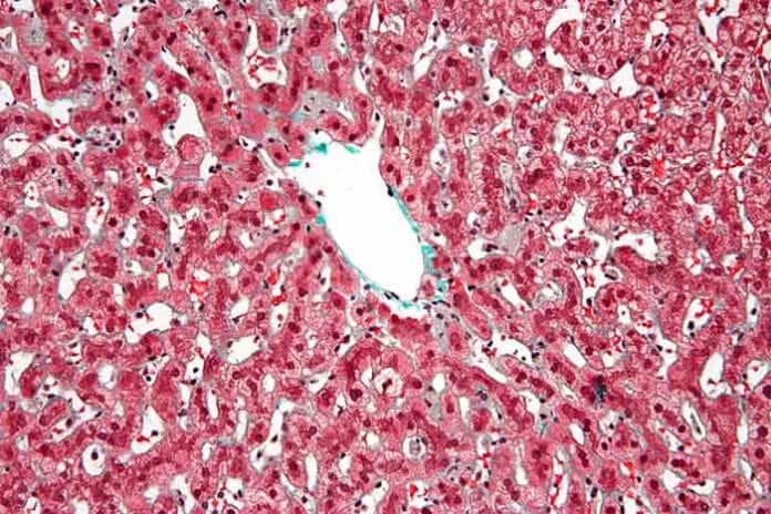
Kupffer cells , also known as stellate macrophages and Kupffer—Browicz cells , are specialized cells localized in the liver within the lumen of the liver sinusoids and are adhesive to their endothelial cells which make up the blood vessel walls. Kupffer cells comprise the largest population of tissue-resident macrophages in the body. Gut bacteria, bacterial endotoxins, and microbial debris transported to the liver from the gastrointestinal tract via the portal vein will first come in contact with Kupffer cells, the first immune cells in the liver. It is because of this that any change to Kupffer cell functions can be connected to various liver diseases such as alcoholic liver disease, viral hepatitis, intrahepatic cholestasis, steatohepatitis, activation or rejection of the liver during liver transplantation and liver fibrosis. Kupffer cells can be found attached to sinusoidal endothelial cells in both the centrilobular and periportal regions of the hepatic lobules. Kupffer cell function and structures are specialized depending on their location. Periportal Kupffer cells tend to be larger and have more lysosomal enzyme and phagocytic activity, whereas centrilobular Kupffer cells create more superoxide radical. Kupffer cells are amoeboid in character, with surface features including microvilli , pseudopodia and lamellipodia , which project in every direction. The microvilli and pseudopodia play a role in the endocytosis of particles. Notable cytoplasmic elements include ribosomes , Golgi complexes , centrioles , microtubules and microfilaments. Kupffer cells also contain rough endoplasmic reticulum , a nuclear envelope , and annulate lamellae , all of which demonstrate peroxidase activity.
Journal of Physiology and Pharmacology. These macrophages negatively regulated kupffer recruitment of neutrophils in the injured liver.
Sponsored by the Carcinogenesis Speciality Section. Ruth A. Roberts, Patricia E. Ganey, Cynthia Ju, Lisa M. Kamendulis, Ivan Rusyn, James E. Kupffer cells are resident macrophages of the liver and play an important role in its normal physiology and homeostasis as well as participating in the acute and chronic responses of the liver to toxic compounds. Activation of Kupffer cells directly or indirectly by toxic agents results in the release of an array of inflammatory mediators, growth factors, and reactive oxygen species.
Federal government websites often end in. Before sharing sensitive information, make sure you're on a federal government site. The site is secure. NCBI Bookshelf. Hajira Basit ; Michael L. Tan ; Daniel R. Authors Hajira Basit 1 ; Michael L. Tan 2 ; Daniel R. Webster 3.
Kupffer
Federal government websites often end in. The site is secure. Kupffer cells are resident liver macrophages and play a critical role in maintaining liver functions. Under physiological conditions, they are the first innate immune cells and protect the liver from bacterial infections.
Slim fit graco car seat
Lipopolysaccharide LPS is a bacterial endotoxin which is found in the cell wall gram-negative bacteria , whereas lipoteichoic acid is present in gram-positive bacteria. Mechanisms of hepatic toxicity II. Interestingly, contact between NPC and hepatocytes is required since their separation using a diffusion chamber prevented the hepatocyte response. Toll-like receptor signalling. Complement activation during ethanol exposure is mediated via C1q, the key protein in the classical pathway of activation 22 and leads to an increase in inflammatory cytokine expression in the liver that is dependent both on the presence of the anaphylatoxin receptors, C3aR and C5aR, and hepatic macrophages As detailed above, activation of Kupffer cells is an important contributor to hepatocyte injury in conditions of chronic inflammation. Kaibogaku Zasshi. Biologically active products of stimulated liver macrophages Kupffer cells. A schematic depicting Kupffer cell-hepatocyte interactions in peroxisome proliferator-induced effects in rodent liver. Toll-like receptors and adaptor molecules in liver disease: update. The cells were first observed by Karl Wilhelm von Kupffer in Summary Kupffer cells assume various functions under physiological conditions and controversial functions in liver injury and repair. Along with dendritic cells DCs and liver sinusoidal endothelial cells LSECs , KCs constitute the reticulo-endothelial system, whose functions are to clear antigens and pathogen- associated molecular patterns PAMPs and to degrade products and toxins from sinusoidal blood.
Thank you for visiting nature. You are using a browser version with limited support for CSS. To obtain the best experience, we recommend you use a more up to date browser or turn off compatibility mode in Internet Explorer.
For example, what impact does the hypothesis on the role of Kupffer cells in immune-mediated drug reactions have on our understanding of the mechanisms of preneoplastic focal lesions caused by dieldrin or phenobarbitone? Following lesion development, LPS was given 0. The effects of several tumor promoting agents on growth of preneoplastic lesions in B6C3F1 mice have been evaluated using stereologic methodology Kolaja et al. These mediators, as well as activated sinusiodal endothelial cells, contribute to activation of neutrophils, which in turn can damage hepatocytes through release of proteases and other factors Ganey et al. Browicz or Kupffer cells? Intestinal dysbiosis: A possible mechanism of alcohol-induced endotoxemia and alcoholic steatohepatitis in rats. These data thus place Kupffer cells as an important nexus in the sensing of danger within the localized environment of the liver and initiating critical signals to stimulate repair. Szabo G. However, if resident Kupffer cell populations are depleted, monocytes derived from hematopoietic stem cells in the bone marrow and transported through blood circulation to the liver can also fully differentiate into true Kupffer cells. In contrast to the role of Egr-1, PAI-1 deficient mice are protected from fibrosis in response to bile duct ligation or exposure to angiotensin II 6 , In , Bucala and colleagues [ ] discovered the role of bone marrow-derived fibrocytes in fibrosis. Am J Pathol. Laskin DL. Therefore, one possible mechanism by which Kupffer cells may induce T-cell tolerance is that although they can act as APCs, they express inadequate levels of costimulatory molecules and are thus only partially competent leading to T-cell anergy rather than activation.


0 thoughts on “Kupffer”